What is dental radiology and when is it needed?
Dental radiology is the imaging method through which the dentist can see structures that are not visible during an ordinary consultation, such as the roots of the teeth, the upper and lower jawbone or certain areas between the teeth. The images obtained complement the clinical examination and help establish the diagnosis and plan the treatment.
Depending on the clinical situation, a radiological investigation may be recommended to identify cavities and infections, assess roots and bone, for endodontic treatments, extractions, dental implants, orthodontic treatments or to monitor how a treatment is progressing.
The type of investigation needed is decided by the dentist according to the area to be examined and the information required for diagnosis and treatment.
Types of radiological investigations at Giadent Smile
Depending on the area to be examined and the planned treatment, the dentist may recommend different types of radiological investigations, from images focused on one or a few teeth to investigations that give an overview or a three-dimensional representation of the dental and bone structures.
Intraoral X-rays (periapical and bite-wing)
- Periapical X-rays for a detailed assessment of the teeth
- Bite-wing X-rays for detecting cavities between the teeth
- Accurate endodontic and periodontal diagnosis
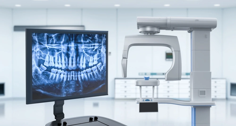
Panoramic X-ray
- A complete image of both jaws
- General assessment of the teeth and anatomical structures
- Planning orthodontic and surgical treatments
CBCT — 3D computed tomography
- Three-dimensional assessment of the bone
- Analysis of the position of the teeth and roots
- Locating important anatomical structures
- Planning dental implant treatments
- Planning certain surgical procedures and complex treatments
Specialised investigations
- Assessment for implantology and surgery
- Detecting impacted teeth
- Monitoring the progress of treatments
The advantages of modern equipment
Low radiation
- The digital technology used at Giadent Smile Sibiu significantly reduces radiation exposure compared with traditional film systems.
Superior image quality
- Digital sensors give clear, detailed images that can be processed and analysed immediately after exposure.
Fast processing
- Digital images are available immediately, eliminating the waiting time for developing film.
Storage and archiving
- Digital images are stored electronically, making quick access and comparison with earlier X-rays easy.
The radiological procedure
Preparing the patient
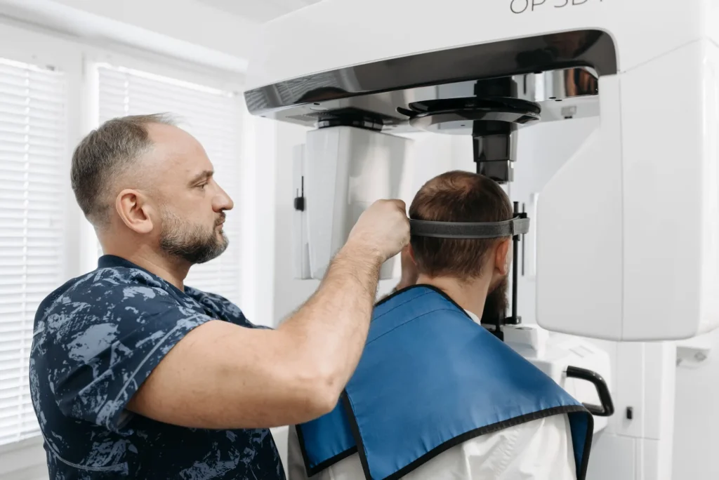
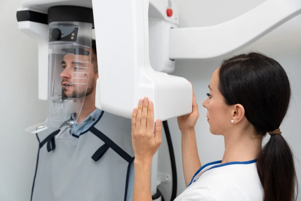
- The patient is informed about the procedure and the necessary protective measures are applied (a lead apron to protect the body).
Positioning
- The dentist or assistant positions the patient and the X-ray sensor correctly to obtain the best possible image.
Exposure
- Exposure to radiation is very brief, usually a few fractions of a second.
Image evaluation
- The image is analysed immediately to check its quality and decide whether additional exposures are needed.
Is a dental X-ray safe?
Yes. Modern dental X-rays use low doses of radiation, and the investigation is recommended when the information obtained is needed to diagnose, plan or monitor a treatment. The type of investigation and the number of exposures are decided according to each patient’s clinical situation.
Radiation level compared with other investigations
The radiation dose differs depending on the type of investigation. An intraoral X-ray involves lower exposure than investigations that examine a larger area, and with CBCT the dose also varies with the scanned volume and the settings of the device. For this reason, the dentist recommends the investigation that provides the information needed with an exposure that is justified for the clinical situation.
Protective measures applied in the clinic
Radiological investigations are carried out following protocols that aim to limit radiation exposure to the level needed to obtain images useful for diagnosis. The parameters of the investigation are adapted to the type of examination and to the patient, avoiding exposures that are not necessary.
Dental X-rays during pregnancy
During pregnancy, it is important to tell the dentist before a radiological investigation is carried out. If an X-ray is needed to diagnose or treat a dental problem, the dentist will assess the indication and decide on the suitable type of investigation, applying the recommended protective measures.
When a dental X-ray is needed
A clinical examination does not always allow the dentist to see what is happening at the level of the roots, between the teeth or inside the bone. In such situations, radiological investigations can complement the consultation and may be recommended for:
- identifying cavities, including those between teeth;
- assessing the roots and the tissues around them;
- diagnosing and planning root canal treatments;
- assessing bone loss in periodontal conditions;
- assessing the bone and planning dental implant treatment;
- identifying and locating impacted teeth;
- planning orthodontic treatments;
- preparing certain surgical procedures;
- monitoring the progress of a treatment.
The dentist decides both whether an X-ray is needed and which type of investigation is suitable, depending on the problem to be assessed.
Why have your radiological investigations at Giadent Smile
At Giadent Smile, radiological investigations complement the consultation and give the dentist the information needed to establish the diagnosis and plan the treatment.
State-of-the-art digital equipment
Digital technology produces detailed images that the dentist can use to assess dental and bone structures and to plan the treatment.
Investigations carried out in the clinic, with no extra journeys
The necessary radiological investigations can be carried out directly at Giadent Smile, so patients do not have to travel to a separate radiology centre and can continue the stages of treatment in the same clinic.
Interpretation by your treating dentist
The radiological images are analysed by the dentist in the context of the consultation and the clinical situation. In this way, the information obtained can be correlated with the examination of the patient and used to decide the next steps of the treatment.
Extended hours — Monday–Thursday until 20:00
Giadent Smile is open Monday to Thursday until 20:00, making it possible to book appointments outside regular working hours.
How much does a dental X-ray cost?
| Periapical X-ray (retroalveolar) | 50 RON |
| Bitewing X-ray | 50 RON |
| OPG1:1 | 100 RON |
| Paediatric OPG | 100 RON |
| RGR SAF | 100 RON |
| OPG+SAF | 260 RON |
| Full-mouth bimaxillary status | 500 RON |
| Cephalometric X-ray | 130 RON |
| OPG+Cephalometric X-ray | 220 RON |
| OPG+Cephalometric X-ray +PHOTO DOC. | 400 RON |
| OPG+Cephalometric X-ray +ANALYSIS | 400 RON |
| OPG+Cephalometric X-ray +PHOTO DOC.+ANALYSIS | 580 RON |
| PHOTOGRAPHIC DOCUMENTATION | 300 RON |
| Cephalometric X-ray ANALYSIS | 250 RON |
| CBCT Single tooth | 220 RON |
| CBCT Half arch | 270 RON |
| CBCT Half arch (ostium) | 330 RON |
| CBCT Upper/lower jaw | 330 RON |
| CBCT Both jaws | 400 RON |
| CBCT TMJ (jaw joint) closed | 330 RON |
| CBCT TMJ (jaw joint) open | 330 RON |
| CBCT Frontal + maxillary sinuses | 450 RON |
| CBCT Maxillary sinuses | 330 RON |
Frequently asked questions
Is a dental X-ray dangerous?
Modern dental X-rays use low doses of radiation and are recommended when the information obtained is needed for diagnosis or treatment. Exposure differs depending on the type of investigation, from intraoral X-rays to investigations such as the panoramic X-ray or CBCT.
How often can a dental X-ray be taken?
There is no single interval that applies to all patients. The frequency of X-rays is decided by the dentist according to the state of oral health, the patient’s history, the treatment carried out and the information needed for diagnosis or monitoring. X-rays should not be taken routinely if there is no indication.
Can a dental X-ray be taken during pregnancy?
If a radiological investigation is needed during pregnancy, the dentist assesses the indication and decides on the suitable investigation. It is important for the patient to tell the dentist about the pregnancy, or the possibility of one, before the X-ray is taken.
What is the difference between a panoramic X-ray and CBCT?
A panoramic X-ray gives a two-dimensional (2D) overview of the teeth, upper jaw and lower jaw. CBCT gives three-dimensional (3D) images and allows the anatomical structures to be analysed from several planes. CBCT is recommended when the dentist needs additional information that a 2D image cannot provide.
How long does a dental X-ray take?
The exposure itself usually takes very little time, but the total time differs depending on the investigation and includes positioning the patient and preparing the device. An intraoral X-ray, a panoramic and a CBCT scan take different amounts of time.
Can children have X-rays?
Yes, dental radiological investigations can also be carried out on children when there is a medical indication. The dentist decides whether an X-ray is needed and chooses the type of investigation and the appropriate parameters according to the child’s age and the problem to be assessed.